
Project
VR Anatomy Atlas
A virtual reality software for teaching human anatomy: over 360 organs that can be grabbed, cut, removed, and re-assembled in a virtual operating room.
01Overview
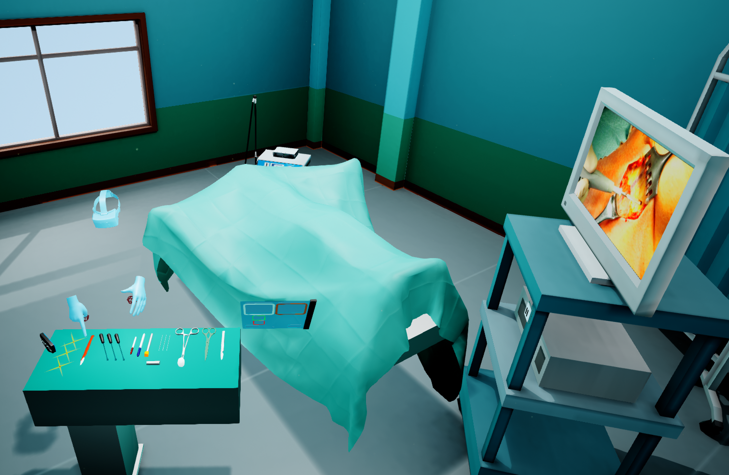
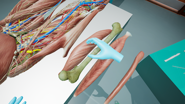
The Immersive Anatomy Atlas transports users into a realistic virtual operating room that contains an anatomical model, instruments, and useful tools. With the help of virtual hands, 360 organs can be grasped to study them closely and to learn their Latin names — for example by removing a heart from the body and inspecting it from every angle.
The software is used both for free exploration and for targeted teaching: teachers can prepare specific scenarios, such as thyroid surgery, and challenge students with them later. It is developed at the CGVR lab, University of Bremen, in cooperation with the Pius-Hospital Oldenburg, and is available in German and English.
Part of the CGVR research program at the University of Bremen: project page at Uni Bremen →
02Videos
03The anatomical model
The model contains over 360 detailed organs — muscles, bones, nerves, and the lymphatic system. Organs can be freely removed, examined, and re-attached: if the user moves an organ roughly back to its original position, it snaps perfectly into place. Every organ is tagged with its Latin name, which is shown when interacting with it, and parts of the body can be hidden to show, for example, only the skeletal system.
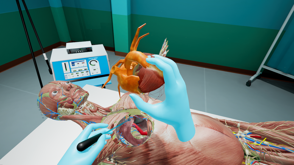
Grab any organ
Organs can be freely removed from the model and closely examined, in either hand.
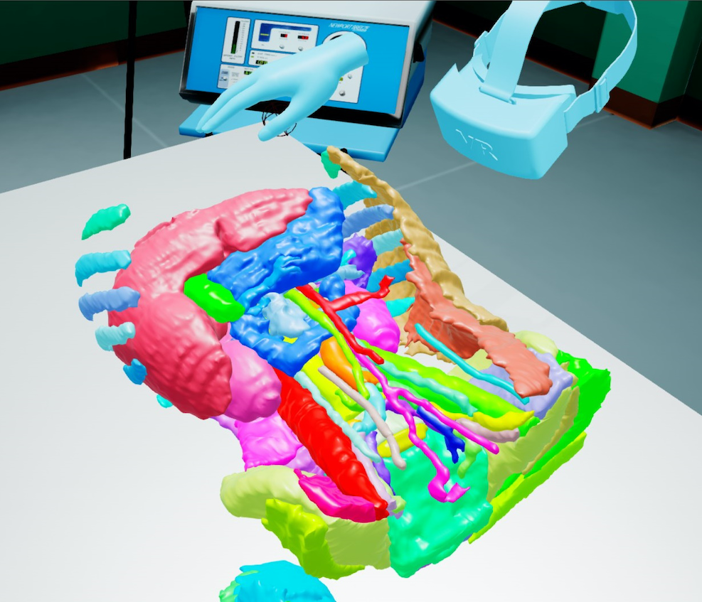
Alternate views
Includes a modified version of the SPL Abdominal Atlas and customizable visibility per body system.
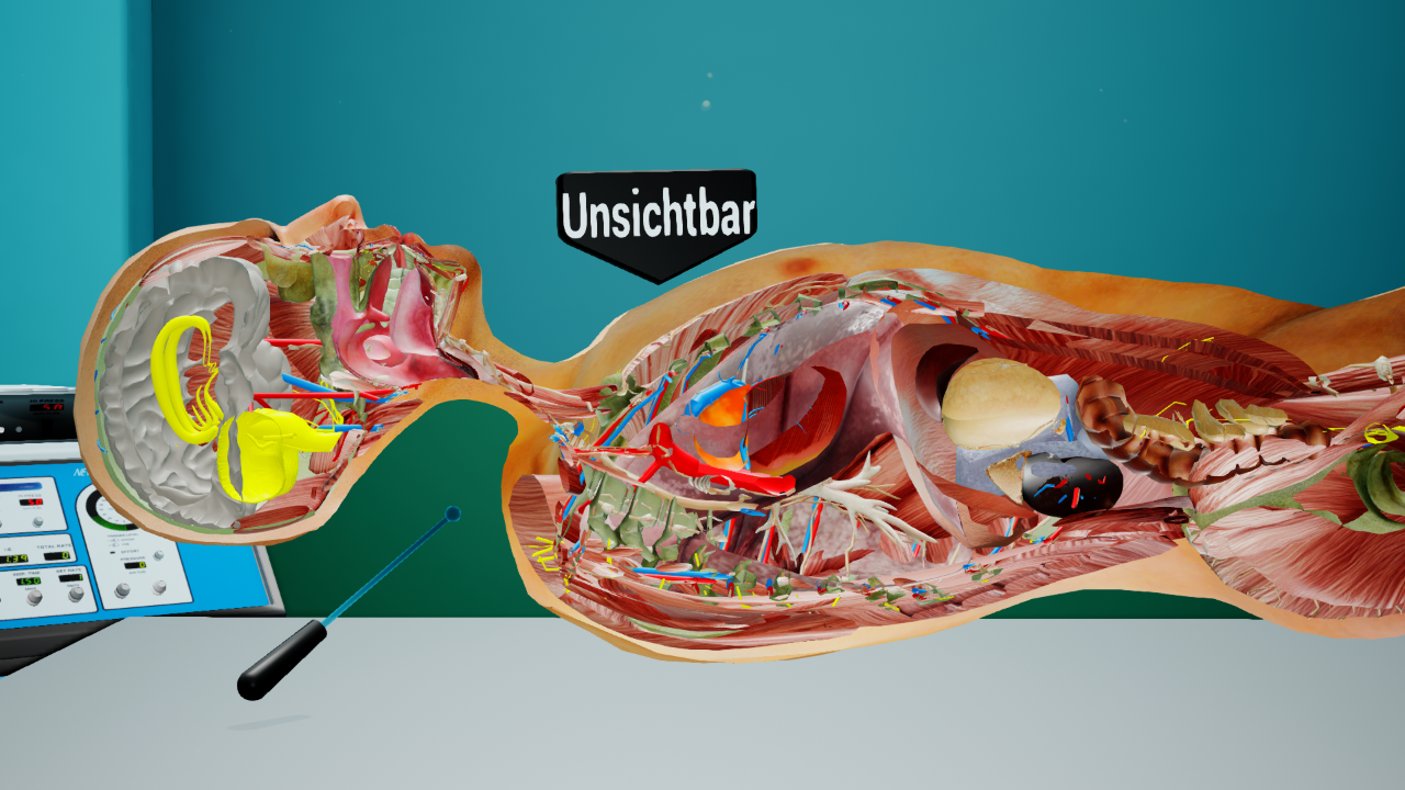
Real-time cross-sections
Virtual tools create cross-sections of any organ to reveal what is inside.
04Tools and instruments
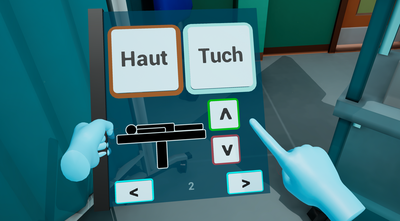
- Cross-sections can be created simply by moving the appropriate tool into the desired place.
- The cloth covering the anatomical model can be cut with a tool representing scissors.
- Organs can be marked, causing them to bleed when touched with a scalpel; another tool stops the bleeding.
- A laser pointer, marker pen, and eraser support visualization and preparation.
- Target positions can be defined; students hit them with a needle and their precision is displayed on-screen.
- Users can use up to two tools at once — one in each hand.
05The operating room
The model lies on an operating table in a fully equipped virtual operating room, complete with ceiling lights that can be dimmed and an ambient soundscape. A video player in the room can show real-life operations in VR. A virtual camera controlled with mouse and keyboard lets spectators and teachers look over the user's shoulder, and organ and tool arrangements can be saved and loaded to teach scenarios to different students.
06Gallery


07Try it
The software is free to download: ImmersiveAnatomy.zip and a brief manual. Requirements: Windows 10/11 64-bit, SteamVR, and a SteamVR-capable headset (e.g. HTC Vive, Meta Quest 3). The software is licensed under the EUPL v1.2.
08Key Results
- An empirical usability study with medical students showed significantly higher satisfaction for the VR environment compared to classical teaching tools (p = 0.012; Frontiers in Surgery, 2018).
- A follow-up study found significantly better results for the VR group in both immediate and delayed tests of factual medical knowledge (Anatomical Sciences Education, 2021).